
Couinaud classification of hepatic segments Radiology Reference
Segmental anatomy Traditionally, the liver was divided into four anatomical lobes: the right, left, caudate and quadrate lobes.

Diagram and Wiring Diagram Of Liver Segments
They are classically described ('classic lobules') as hexagonal structures made of six vertically aligned portal canals with a central vein. However, microscopic evaluation of the liver usually shows a lack of classic liver lobule as a well-defined connective tissue septum is usually lacking. Liver lobules are enveloped by Glisson's capsule.
Liver Radiology Key
Hepatic segments Diagram Diagrams of the division of the liver into segments. Hepatic sections Diagram Diagrams of the division of the liver into sections. Case Discussion Diagrams of the division of the liver into segments and sections based on the Couinaud classification. 3 articles feature images from this case

Couinaud classification of hepatic segments Radiology Reference
1/4 Synonyms: none Anatomically, the liver is viewed as having four main lobes. There is a smaller left lobe and a larger right lobe (that are separated along the attachment of the falciform ligament ), as well as a caudate and a quadrate lobe (which are part of the anatomical right lobe).

Couinaud classification of hepatic segments Radiology Reference
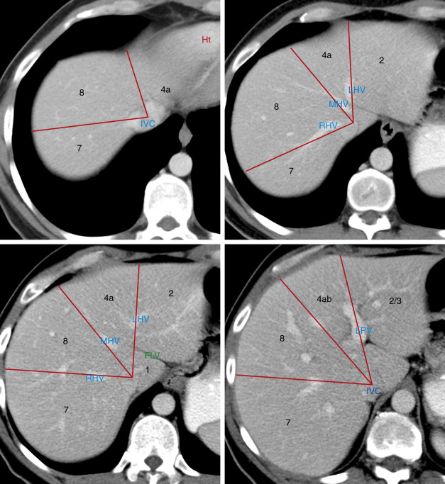
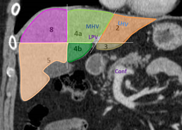
In a portal venous phase CT, the right, middle, and left hepatic veins are usually filled with contrast-mixed blood and can be seen emptying into the inferior vena cava, which is found along the back side of the liver. The portal and hepatic veins divide the liver into segmental anatomy that allows radiologists and surgeons to be very specific.

bismuth of liver Google Search Radiology, Ultrasound, Liver anatomy
Citation, DOI, disclosures and article data. The AAST (American Association for the Surgery of Trauma) liver injury scale, recently revised in 2018, is the most widely used liver injury grading system 3. The 2018 update incorporates "vascular injury" (i.e. pseudoaneurysm, arteriovenous fistula) into the imaging criteria for visceral injury 3.

Diagram Of Liver Human Liver With Labels Illustration Stock Photo Alamy
The hepatic segmentation (lobes, parts, divisions and segments) is the oganization of the liver into parts, divisions and segments.

Anatomy of the liver segments Liver anatomy, Diagnostic medical
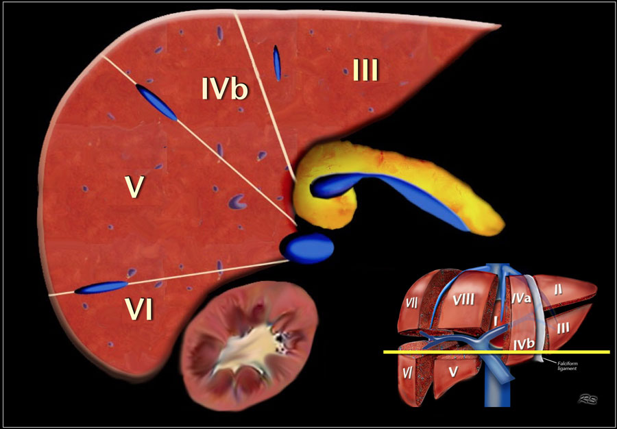
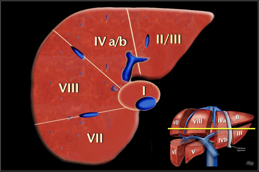
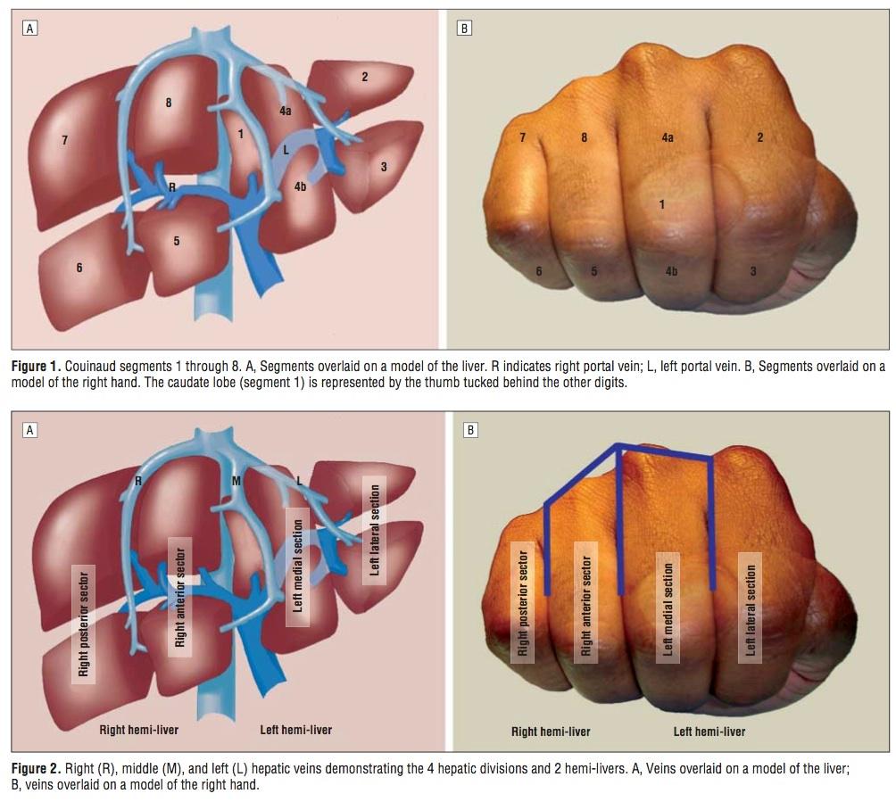
Segmental anatomy Segmental anatomy according to Couinaud. Click to enlarge. Couinaud classification The Couinaud classification of liver anatomy divides the liver into eight functionally indepedent segments. Each segment has its own vascular inflow, outflow and biliary drainage.
Liver Ultrasound Anatomy Anatomical Charts & Posters
The segments are represented by the following: segment 1: (caudate): the thumb, in the palm of the hand segment 2: index finger proximal phalanx segment 3: index finger middle phalanx segment 4a: middle finger proximal phalanx segment 4b: middle finger middle phalanx segment 5: ring finger middle phalanx segment 6: little finger middle phalanx

Couinaud classification of hepatic segments Radiology Reference
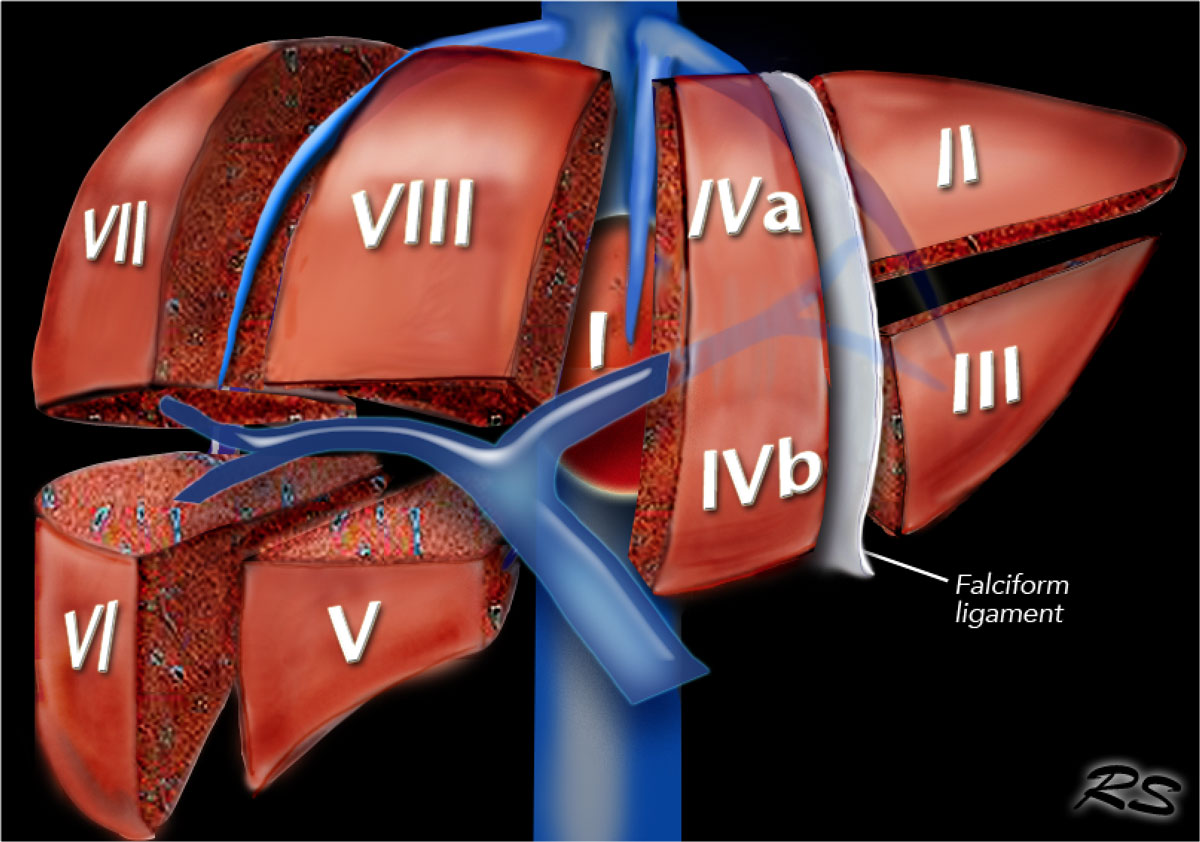
A liver segment is one of eight segments of the liver as described in the widely used Couinaud classification (named after Claude Couinaud) in the anatomy of the liver.This system divides the lobes of the liver into eight segments based on a transverse plane through the bifurcation of the main portal vein, arranged in a clockwise manner starting from the caudate lobe.
Liver Anatomy Segments
It is the preferred anatomy classification system as it divides the liver into eight independent functional units (termed segments) rather than relying o. Playlist Liver Segments (Coronal) 1 case No description provided Playlist Liver Segments (Axial) 1 case No description provided Case Couinaud liver segments (illustration)
Сегменты печени на узи схема Медицинский справочник lklinika.ru
Future liver volume prior to major hepatectomy. Liver volumetry is indicated in patients undergoing major hepatic resection [], defined as resection of four or more segments according to the Couinaud classification [].Most hepatectomies are performed for the treatment of neoplasms, including primary liver cancer (e.g. hepatocellular carcinoma and cholangiocarcinoma), liver metastases (e.g.
segmental anatomy of the liver
Liver segmentation for volumetric assessment is indicated prior to major hepatectomy, portal vein embolisation, associating liver partition and portal vein ligation for staged hepatectomy (ALPPS) and transplant. Segmentation software can be categorised according to amount of user input involved: manual, semi-automated and fully automated.
Liver Segments Ct Scan
PURPOSE: To evaluate qualitatively and quantitatively the current procedures for radiologic delineation of the segmental and subsegmental anatomy of the liver. MATERIALS AND METHODS: Vascular casts of 10 livers were examined with helical computed tomography (CT). Liver segmental and subsegmental anatomy were determined on the CT scans according to customary radiologic practice guidelines. CT.

From the Angio Suite to the γCamera Vascular Mapping and 99mTcMAA
The left hepatic artery runs vertically towards the umbilical fissure and supplies segments 1, 2 and 3. It usually gives off a middle hepatic artery branch that runs towards the right side of the umbilical fissure and supplies segments 4a and 4b 2 . Within the liver, the left hepatic artery divides into: medial segmental branch.
The Radiology Assistant Liver Segmental anatomy
The middle hepatic vein separates the right liver from the left liver and the right hepatic vein separates the right posterior sector and anterior sector. The plane of the portal vein is used to divide the different segments into upper and lower sectors (this plane is correct for the segments of the right liver but false for segments II and III.